
Treatment(Proton Therapy)
- HOME
- Cancer(Treatment)
- Treatment(Proton Therapy)
- Understanding Proton Beam Therapy for Head and Neck Cancer Through Real Cases: Tongue Cancer, Floor of Mouth Cancer, and Adenoid Cystic Carcinoma ~ Southern TOHOKU Proton Therapy Center
Understanding Proton Beam Therapy for Head and Neck Cancer Through Real Cases: Tongue Cancer, Floor of Mouth Cancer, and Adenoid Cystic Carcinoma ~ Southern TOHOKU Proton Therapy Center
26.10.23
What do we hope for when we are diagnosed with head and neck cancer ?
Not only successful treatment, but also the chance to regain our quality of life and sense of self after treatment.
We introduce a treatment approach centered on proton therapy—a path toward regaining the joy of eating, the sense of taste, and the freedom to speak.
These clinical cases were provided by the Southern TOHOKU Proton Therapy Center, whose treatment achievements were highlighted in the Nikkei series “Given One Year to Live: A Patient with Maxillary Sinus Cancer Entrusts His Life to the Words ‘You’ll Be Fine’,” which began on August 29, 2026.
Here, you’ll find insights to help you envision the future for yourself and your loved ones.
Case 1

4 40-year-old Female
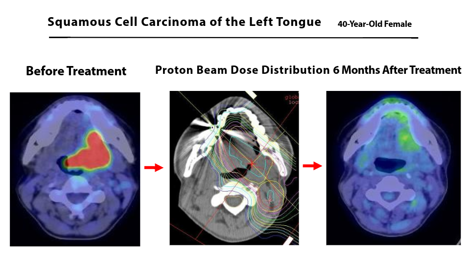
- Tongue Cancer (Left-sided Tongue Squamous Cell Carcinoma)
- A Case in Which the Patient Declined Surgery and Chose Proton Beam Therapy Combined with Intra-Arterial Chemotherapy
Diagnosis
・Left-sided tongue squamous cell carcinoma
・cT4aN1M0
TNM classification indicating the extent of cancer progression.
The tumor had spread into surrounding tissues and metastasized to cervical lymph nodes, but no distant metastasis was detected.
Path to Diagnosis
The patient sought medical attention because of persistent oral ulcers and was diagnosed with tongue cancer.
Because she did not wish to undergo surgery with reconstructive procedures, she was treated with a combination of systemic chemotherapy, radiation therapy, proton beam therapy, and intra-arterial chemotherapy.
Treatment Details
Total radiation dose: 69 Gy
・Systemic chemotherapy (5-FU + cisplatin)
・X-ray radiotherapy: 36 Gy / 20 fractions
・Proton beam therapy: 33 Gy(RBE) / 15 fractions
・Intra-arterial chemotherapy (cisplatin): 5 sessions
※Gy is a unit of radiation dose that indicates the amount of radiation energy absorbed by body tissues.
During Treatment
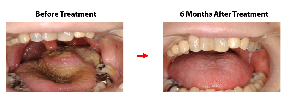
・Mucositis
・Temporary dysphagia
・Taste disturbance
Because of these treatment-related effects, a PEG tube was temporarily placed. She regained the ability to eat orally two months after treatment, and the PEG tube was removed six months after treatment.
After Treatment
- PET imaging performed 6 months after treatment showed complete disappearance of FDG uptake.
- Oral intake was resumed 2 months after treatment
- The patient has remained free of recurrence for more than 10 years following treatment.
Case 2
66-year-old Male (Wine Sommelier)
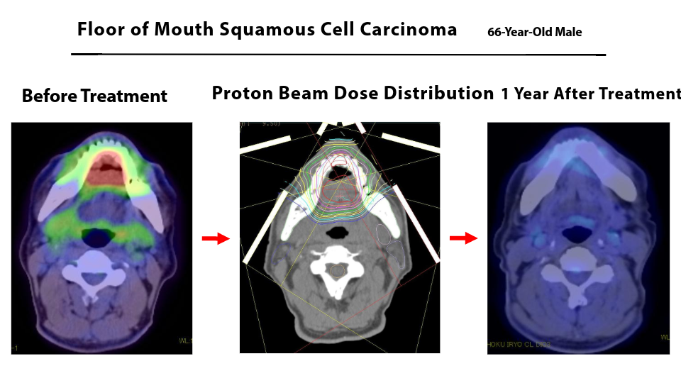
- Floor of Mouth Cancer (Squamous Cell Carcinoma of the Floor of the Mouth)
- A Treatment Case Chosen by a Patient Who Prioritized the Preservation of Oral Function
・Squamous cell carcinoma of the floor of the mouth
・cT4aN2cM0
The cancer had spread to surrounding tissues and to lymph nodes on both sides of the neck, but no distant metastasis was detected.
Path to Diagnosis
The patient presented with a tumor in the floor of the mouth and swallowing difficulties and was diagnosed at a university hospital.
Because preserving oral function was important for his profession, he chose a treatment approach combining radiation therapy, proton beam therapy, and intra-arterial chemotherapy as an alternative to surgery.
Treatment Details
Total radiation dose: 66 Gy
・Intensity-Modulated Radiation Therapy (IMRT): 40 Gy / 20 fractions
・Proton beam therapy: 26 Gy(RBE) / 13 fractions
・Intra-arterial chemotherapy (cisplatin): 4 sessions
During Treatment
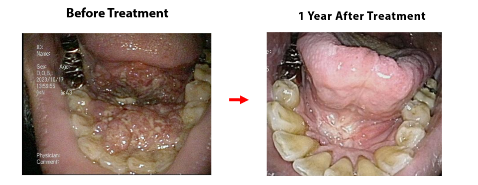
・Mucositis
・Temporary taste disturbance
Although these symptoms occurred, oral intake remained possible throughout treatment.
After Treatment
- Favorable local tumor control was confirmed 1 year after treatment
- The patient returned to normal daily activities and was able to continue working as a chef. However, continued alcohol consumption was considered a contributing factor, and a new primary tongue cancer developed during follow-up 3 years after treatment. The lesion was detected at an early stage and was treated surgically.
Case 3
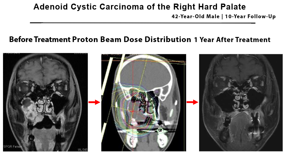
42-year-old Male
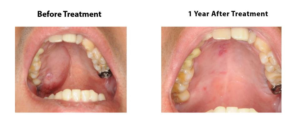
- Adenoid Cystic Carcinoma of the Right Hard Palate
- A Case of Proton Beam Therapy for a Tumor Extending to the Skull Base
Diagnosis
・Adenoid cystic carcinoma of the right hard palate
・cT4bN0M0
Advanced hard palate cancer extending to the skull base, without lymph node or distant metastasis.
Path to Diagnosis
The patient presented with swelling of the upper palate and was diagnosed with palatal adenoid cystic carcinoma.
Because the tumor had extended to the skull base and was considered unsuitable for surgical resection, combined treatment with proton beam therapy and intra-arterial chemotherapy was performed.
Treatment Details
・Proton beam therapy: 70.4 Gy(RBE) / 32 fractions
・Intra-arterial chemotherapy (cisplatin): 6 sessions
・Catheter placement through the superficial temporal artery
After Treatment
- Tumor regression was confirmed 1 year after treatment
- Although bilateral lung metastases developed, both lesions were successfully controlled with proton beam therapy.
- Radiation-related brain necrosis was observed 5 years after treatment, but the condition subsequently stabilized, and 10 years have now passed since treatment.
Each patient has unique priorities and expectations for life after treatment.
Treatment-related side effects and long-term outcomes can vary considerably, and not all patients will experience the same results. Nevertheless, as illustrated by the cases presented here, a combination of proton beam therapy and intra-arterial chemotherapy may be a treatment option for selected patients, depending on their medical condition and personal preferences.
We hope that these cases will provide useful insights and serve as a helpful reference for patients and their families as they consider their treatment options.